
重症の肉離れの受傷直後から完治までのエコー画像観察です。
特に血腫の変化や再生の具合が詳しく観察できました。
症例はテニスをされる女性でテニスプレイ中にボレーで左に踏み込んだ際に右脹脛に筋肉が切れた感じと激痛で運動不能となりました。
重症の筋断裂です。
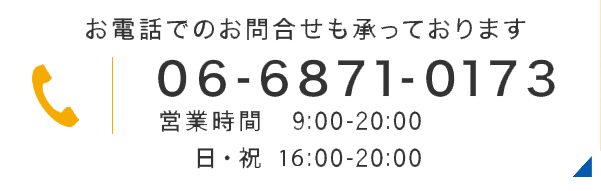
写真①1/18
テニスで負傷。受傷1時間程度で来院。腓腹筋の筋腹に直径2〜3cmの大きな血腫。特に受傷直後の血腫は白い塊状に見えることがある。
このように受傷直後から大きな血腫が見られるものは断裂でも重症である。
踵を接地できず歩行はできない。僅かにも足関節を背屈することができない。
初回の処置は、筋.筋膜の整復とヒールリフト(1.5cm)。電療はエレサスのモード2で2000hz。固定は全くなし。 筋.筋膜の整復で直後から強い痛みと引きつりが減少し歩行可能になった。
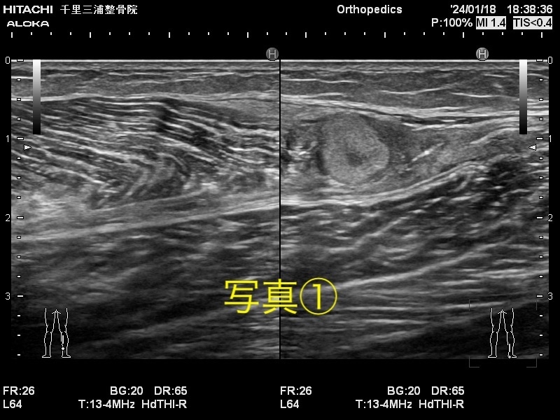
写真② 1/19
血腫が白色から黒色に変化。通常、血腫はこのようにローエコーで見られる。腓腹筋は全体的に腫脹している。しかし腫脹の大きさはかなり抑えられていると思われる。テーピングや固定しなくても腫脹はかなり抑えられる。これはヒールリフトの影響が大きく、踵を持ち上げ、下腿部の筋肉に弛みを与えて患部の負担を軽減している。この時点で破行は伴うが歩行が可能。
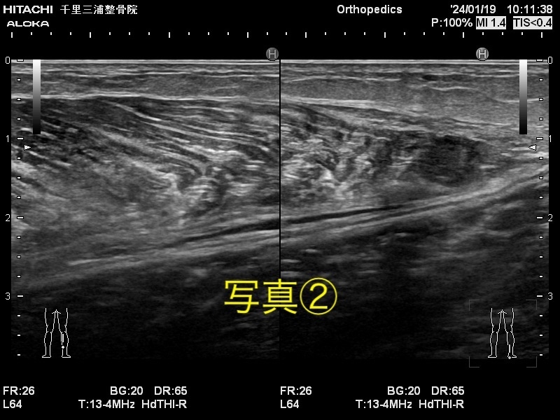
写真③2/2
腓腹筋とヒラメ筋との間に血腫が見られる。これは既に血液のようでなく血漿に近いものと思われる。損傷してから約2週間後によく見られ、血腫が増加しているが、悪化しているわけではない。この時のこの患者さんは患部に痛みが全くなく正常に歩行できる。テニスもある程度可能なレベルに回復している。
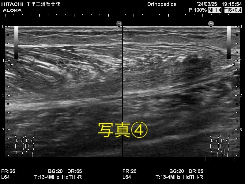
写真④ 3/25
全く別のケガで来院時に撮影。
テニスの試合も参加し、殆ど不安なくプレイしているとの報告あり。
腓腹筋とヒラメ筋の間に明確な筋膜が再生している事が確認できる。当然、血腫は全く見られない。
当院では受傷直後からでも積極的な筋膜の整復を行います。しかしエコーで損傷部の観察は重要で十分に安全を考慮しなければなりません。早期整復はとても重要です。早く回復するだけでなく、後遺症のリスクを大きく下げます。
~english ver~
This is an echo image observation from the immediate onset to complete recovery of a severe muscle tear. We were able to observe in detail the changes in hematoma and the progress of regeneration, particularly. The case involves a female tennis player who experienced a severe muscle tear in her right calf when stepping to the left during a volley while playing tennis, causing intense pain and immobility. It's a severe muscle rupture.
Photo 1/18 Injured during tennis. Visited about an hour after the injury. A large hematoma with a diameter of 2-3 cm in the belly of the calf muscle. Hematomas immediately after the injury may appear as white clumps. Instances where large hematomas are observed immediately after injury indicate severe damage, even if there is no rupture. Unable to walk without touching the heel to the ground. Unable to slightly dorsiflex the ankle. The initial treatment involved realignment of the muscle and fascia and a 1.5cm heel lift. Electrotherapy at 2000hz using the Eleasys mode 2. No immobilization. Immediate reduction in strong pain and spasms after realignment of muscle and fascia, enabling walking.
Photo 2 1/19 Hematoma has changed from white to black. Hematomas are usually seen like this on low echo. The calf muscle is overall swollen. However, the swelling seems to be significantly reduced. Swelling can be significantly reduced without taping or immobilization. This is largely due to the effect of the heel lift, which lifts the heel and relaxes the calf muscles, reducing the strain on the affected area. At this point, walking is possible despite the tear.
Photo 3 2/2 A hematoma is seen between the calf muscle and the soleus muscle. This is already closer to plasma than blood. It is often observed around 2 weeks after injury, with an increase in hematoma, but without worsening. At this point, this patient has no pain in the affected area and can walk normally. Tennis is also somewhat possible at this level of recovery.
Photo 4 3/25 Taken at the time of visit for a completely different injury. Reportedly participating in tennis matches and playing without much anxiety. Clear fascia regeneration between the calf muscle and the soleus muscle can be confirmed. Naturally, no hematoma is visible.
At our clinic, we perform active realignment of the fascia even immediately after injury. However, observing the injured area with ultrasound is important and must be done safely. Early realignment is very important. It not only speeds up recovery but also significantly reduces the risk of sequelae.