
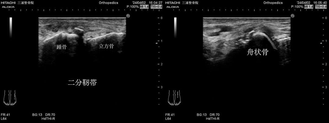
今回は、足首捻挫による二分靭帯損傷および、踵骨前方突起と舟状骨の二箇所の剥離骨折の疑いが見られる症例です。 病院を進めましたが、経過も良好なためこのままここで経過観察をすることにしました。
バレーボールをする大学生の男性。
味方の足を踏み捻挫してしまい、直後から歩行不能となりました。
受傷当日から動けるところまでの数日間、エコー観察ができたので報告します。
病院ではギプスで固定をして、数ヶ月安静と松葉杖が必要になる重症な捻挫になります。
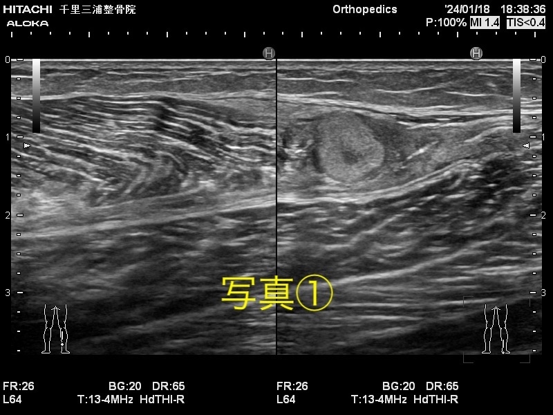
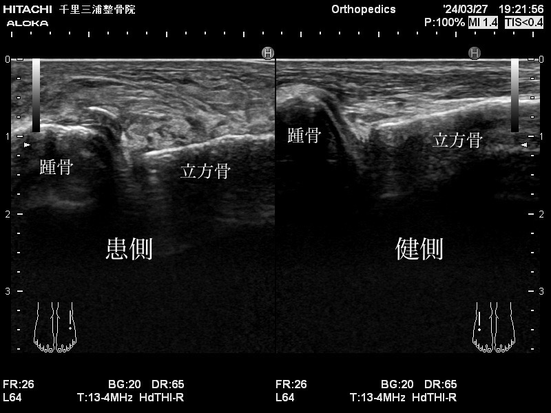
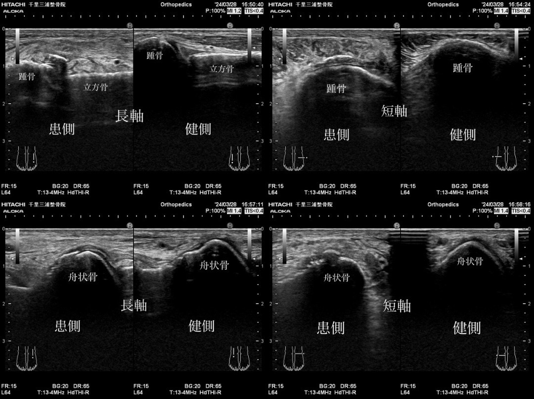
3/27 負傷当日
バレーボールの試合中、バックアタックの着地で味方の足を踏み、捻挫。直後から歩行不能。負傷から数時間後に来院。
エコーで画像では、二分靭帯損傷、踵骨前方突起剥離骨折と舟状骨骨折の疑いが見られる。
底背屈制限があり、足をついて歩行不能。
この日の処置は、徒手整復とヒールリフト(1.5cm)。電療はエレサスのモード1で2,000Hzとオステオトロンを骨折が疑われる箇所に1000Hzで60mW/㎠で20分間した。
固定は一切なし。
徒手整復で跛行はあるが歩行可能。足をつけているが、念の為、松葉杖を貸した。
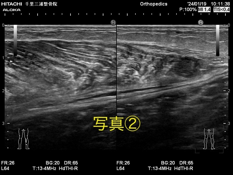
3/28 負傷2日目
腫れは増加しているが、歩行はなんとか可能。
ヒールリフトを着用するとより楽になる。
二分靭帯損傷は、爪先立ちで痛みが出る箇所である。なので、ヒールリフトをすることで踵が持ち上げられ、爪先立ちに掛かる負担が軽減され、歩行時痛が軽減されたと考えられる。
3/29 負傷3日目
腫れの増加なし。
3日目にして痛みがそれほどなく、歩行、ジョギング、軽いジャンプが可能。
3/30 負傷4日目
腫れが減少。
おおよそ、痛めて72時間が炎症の時期です。ここを超えてくると問題なくいけば、自然と腫れは引いてくる。
3/31 負傷5日目
ランニングを15分ほどしたが、痛みなし。
腫れの増加は見られなかった。
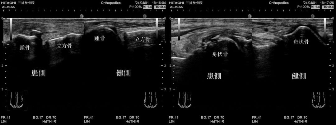
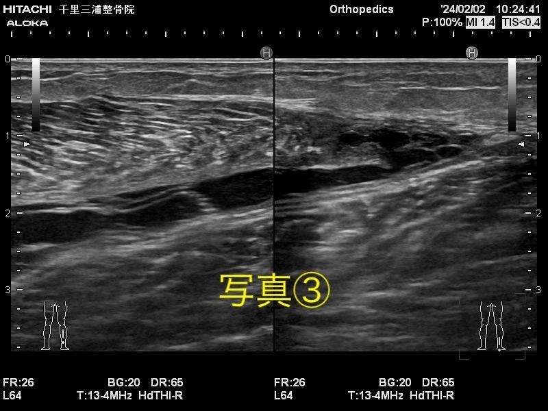
4/1 負傷6日目
ランニングを15分ほどした。つま先の方に痛みが出たきたが、エコーでは問題なし。
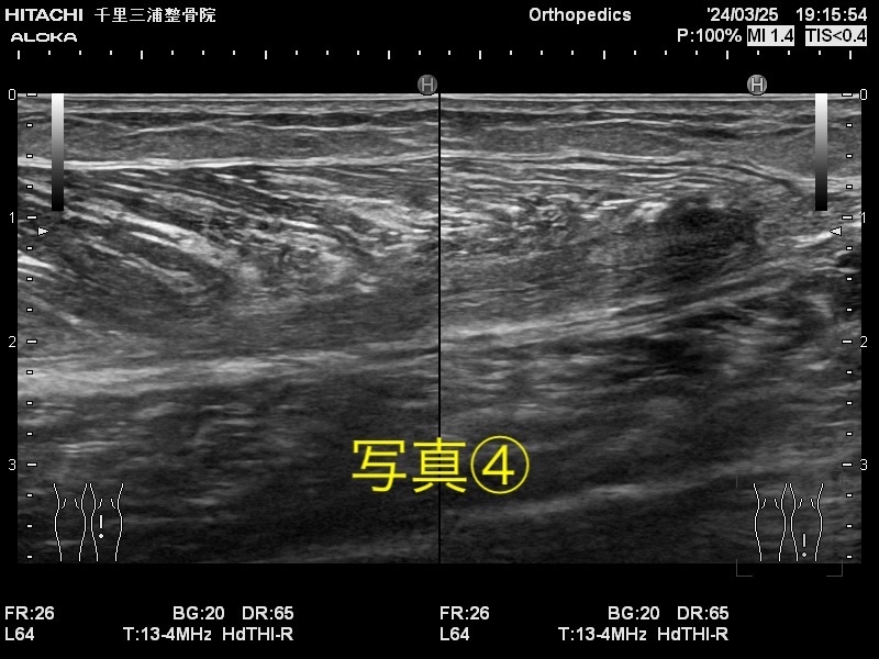
4/2 負傷7日目
腫れが減少。痛みがなく全力でジャンプが可能。
ランニングの時につま先に痛みが残る。
徒手整復で痛み減少。
翌日から練習参加予定。
画像では靭帯損傷がまだまだひどい状態だが、徒手整復で運動は可能になり、今では練習にも復帰しているようです。
怪我をした初日から筋・筋膜の整復をすることで、早期回復の可能性は格段に上がります。
もし怪我をしてしまったら、すぐの来院をおすすめします。
english ver
"Ankle Sprain (Case Suspected of Partial Ligament Tear and Fracture in Two Places)
This case involves a suspicion of a partial ligament tear due to an ankle sprain, as well as suspected fractures in two places: the anterior process of the calcaneus and the avulsion fracture of the navicular bone. Although the patient was advised to proceed with hospitalization, due to a favorable prognosis, it was decided to continue observation here.
The patient is a male college student who plays volleyball.
He sustained the injury by accidentally stepping on a teammate's foot during play, resulting in immediate inability to walk.
Echo observations were possible in the days following the injury, and the following report is based on those observations.
At the hospital, the injury was immobilized with a cast, and it was deemed a severe sprain requiring several months of rest and the use of crutches.
March 27th - Day of Injury
During a volleyball game, the patient stepped on a teammate's foot during a back attack landing, resulting in a sprain. Immediate inability to walk. Arrived at the hospital a few hours after the injury.
On the echo image, there is suspicion of a partial ligament tear, avulsion fracture of the anterior process of the calcaneus, and fracture of the navicular bone.
There is dorsiflexion restriction, and the patient is unable to walk on the affected foot.
Treatment on this day included manual reduction and a 1.5cm heel lift. Electrotherapy was performed using the ElecSAS Mode 1 at 2000Hz and the Osteotron at 1000Hz, 60mW/cm² for 20 minutes in the suspected fracture areas.
No immobilization was applied.
Although limping is present after manual reduction, the patient can walk. Crutches were provided for precautionary measures.
March 28th - Day 2 of Injury
Swelling is increasing but walking is still possible.
Wearing the heel lift provides some relief.
Pain from the partial ligament tear is felt when standing on tiptoes. Therefore, wearing the heel lift raises the heel, reduces the burden on the tiptoes, and alleviates pain during walking.
March 29th - Day 3 of Injury
No increase in swelling.
Pain is not significant on the third day, and walking, jogging, and light jumping are possible.
March 30th - Day 4 of Injury
Swelling is decreasing.
Typically, 72 hours after the injury is the period of inflammation. If this period is surpassed without issues, swelling naturally subsides.
March 31st - Day 5 of Injury
Ran for about 15 minutes without pain.
No increase in swelling observed.
April 1st - Day 6 of Injury
Ran for about 15 minutes. Experienced some pain in the toes, but no issues detected on the echo.
April 2nd - Day 7 of Injury
Swelling is decreasing. Full jumps are possible without pain.
Pain remains in the toes during running.
Pain reduced after manual reduction.
Plan to participate in practice from the next day.
Although the ligament tear is still severe according to the images, exercise is possible after manual reduction, and the patient has returned to practice. Early recovery is greatly enhanced by starting muscle and fascial reduction from the first day of injury. Immediate hospitalization is recommended if an injury occurs."